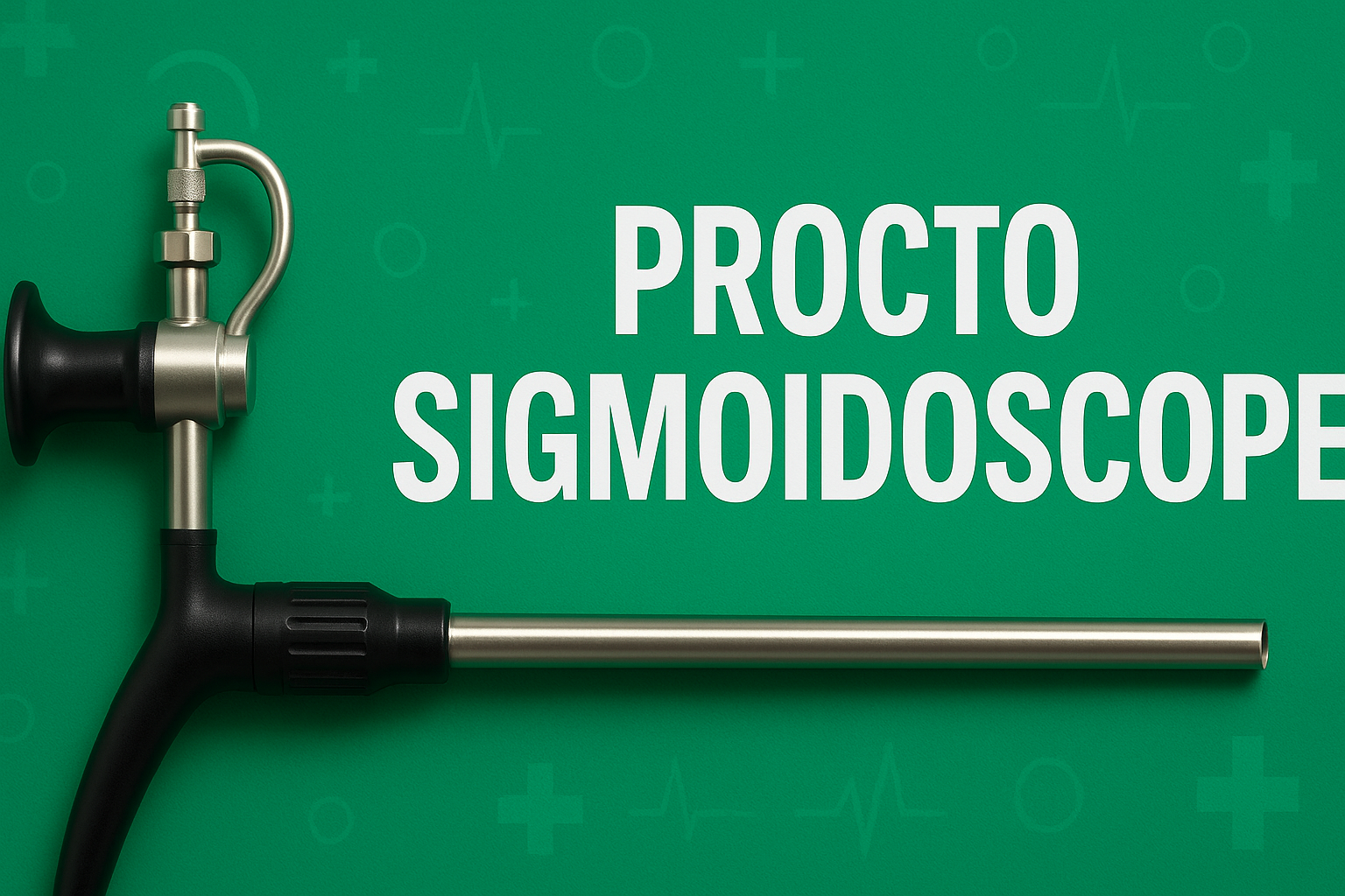
Introduction to Procto Sigmoidoscope Techniques
Procto Sigmoidoscope techniques have become a cornerstone in colorectal screening, offering clinicians a reliable method to detect early mucosal changes in the distal colon. The Procto Sigmoidoscope allows visualization of the sigmoid colon and rectum with greater precision than a simple digital exam. By refining Procto Sigmoidoscope techniques, practitioners can improve polyp detection rates, reduce false negatives, and enhance patient outcomes. In this guide, we’ll explore preparation protocols, visualization strategies, advanced instrumentation, training standards, patient comfort measures, and data management—all aimed at enhancing colorectal screening accuracy with the Procto Sigmoidoscope.
Procto Sigmoidoscope Preparation Protocols
Successful Procto Sigmoidoscope exams start long before the patient arrives in the procedure room. Proper bowel preparation is critical: clear liquid diets for 24 hours, followed by targeted laxative regimens, help ensure mucosal surfaces are free of debris. When scheduling a Procto Sigmoidoscope exam, clinicians should provide clear, written instructions detailing dietary restrictions and timing of oral lavage agents. Adequate hydration must be emphasized to prevent electrolyte imbalances. For patients with diabetes or renal issues, preparation protocols should be individually tailored. By standardizing Procto Sigmoidoscope preparation, teams can minimize cancellations due to poor prep and maximize visualization of the colorectal mucosa.
Optimizing Procto Sigmoidoscope Visualization
Once the bowel is prepared, maximizing visualization during the exam is the next priority. High-definition optics in modern Procto Sigmoidoscope devices provide up to 10× magnification, helping to spot subtle lesions. Adequate insufflation with air or carbon dioxide ensures the lumen is distended uniformly. Gentle patient repositioning—from left lateral to supine—can help flatten sigmoid folds. Dynamic retroflexion in the rectum further reveals hidden areas. Combining white-light endoscopy with narrow-band imaging or chromoendoscopy enhances contrast, making vascular patterns and flat polyps more conspicuous. When implemented consistently, these visualization enhancements in Procto Sigmoidoscope procedures lead to improved lesion detection and fewer missed diagnoses.
Advanced Procto Sigmoidoscope Instrumentation
Technical advances in Procto Sigmoidoscope instrumentation have significantly broadened procedural capabilities. Pediatric-length scopes with slimmer calibers improve comfort, particularly in sensitive patients. Integrated working channels allow simultaneous biopsy and polypectomy without scope withdrawal. Angled-tip optics facilitate easier navigation of redundant sigmoid loops. Some systems now include distal caps that help flatten mucosal folds for panoramic views. Water-immersion techniques, using a controlled stream to lift debris, can replace air insufflation, reducing spasm. When clinicians adopt cutting-edge Procto Sigmoidoscope tools and accessories, they can perform more thorough inspections with fewer interruptions, accelerating throughput and enhancing diagnostic accuracy.
Procto Sigmoidoscope Training and Standardization
High-quality Procto Sigmoidoscope outcomes hinge on consistent training and credentialing. Simulation-based education using virtual-reality scopes allows trainees to develop hand–eye coordination and loop-management skills without patient risk. Procto Sigmoidoscope curricula should include modules on lesion recognition, scope handling, and complication management. Benchmarking performance—such as polyp detection rate, cecal reach (when applicable), and withdrawal time—helps maintain high standards. Regular peer-review sessions and video audits encourage continuous improvement. By establishing standardized Procto Sigmoidoscope training programs and metrics, institutions ensure that all endoscopists meet or exceed quality thresholds for colorectal screening.
Improving Patient Comfort During Procto Sigmoidoscope
Patient tolerance is crucial for effective Procto Sigmoidoscope exams. Adequate analgesia, often with topical lidocaine gel and conscious sedation, can reduce discomfort and encourage deeper inspection. Warmed insufflation gas lessens cramping. Clear, calm communication about expected sensations, along with the option to pause, empowers patients and reduces anxiety. Distraction techniques—such as music or guided breathing—can also improve compliance. In select cases, water-immersion Procto Sigmoidoscope approaches not only enhance visualization but also produce less pain than traditional air methods. By prioritizing patient comfort, clinicians can gather more complete data and reduce repeat examinations due to intolerance.
Data Management in Procto Sigmoidoscope Procedures
Accurate documentation and data analysis play a vital role in Procto Sigmoidoscope quality assurance. Endoscopy reporting software should automatically capture key metrics: date and time, indication, prep quality, depth of insertion, and findings. Standardized photo-documentation protocols ensure that images of suspicious areas are saved in consistent formats. Linking Procto Sigmoidoscope records with pathology systems streamlines tracking of biopsy results. Regular extraction of performance data—such as adenoma detection rate and complication incidence—allows for real-time feedback. When combined with audit cycles, robust data management systems empower teams to identify areas for improvement and maintain high colorectal screening accuracy.
Conclusion: Procto Sigmoidoscope Techniques and Future Directions
As colorectal cancer remains a leading cause of morbidity, refining Procto Sigmoidoscope techniques is more important than ever. Through meticulous preparation protocols, optimized visualization strategies, advanced instrumentation, comprehensive training, patient-centered comfort measures, and rigorous data management, clinicians can significantly boost screening accuracy. Future innovations—such as artificial-intelligence–assisted lesion detection and capsule-based Procto Sigmoidoscope systems—promise to push boundaries even further. By staying abreast of evolving best practices in Procto Sigmoidoscope procedures, healthcare providers can continue to improve early detection rates and save lives through timely intervention.